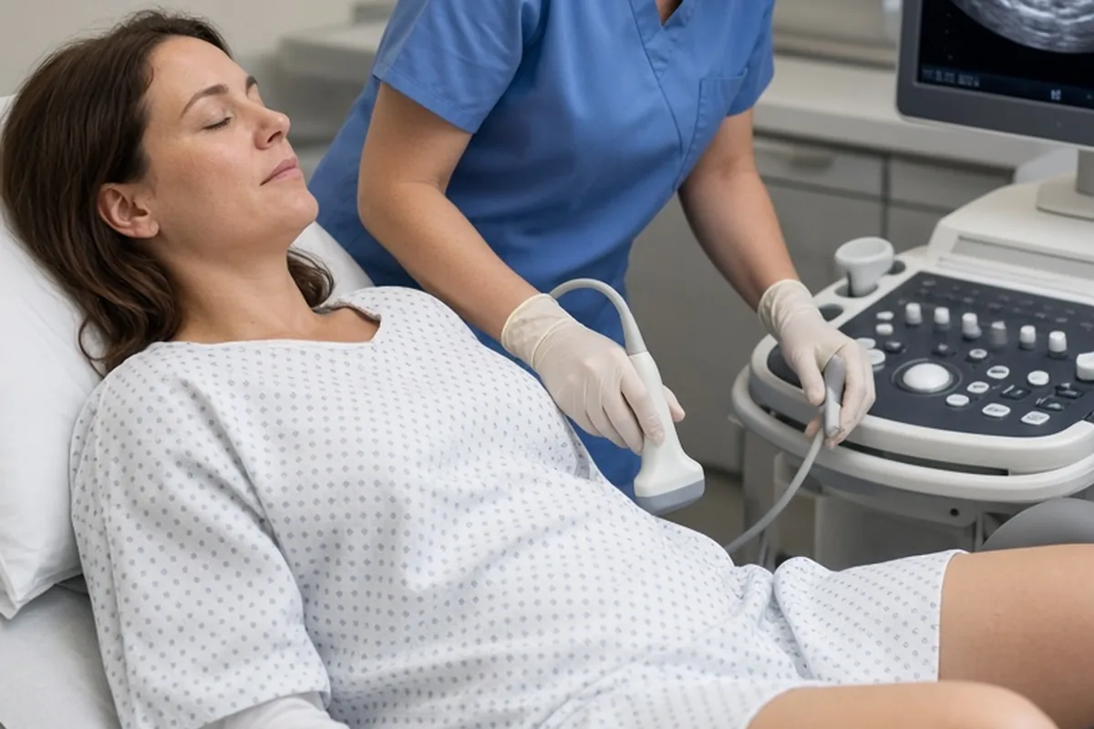
Hydrosalpinx occurs when a fallopian tube becomes blocked and fills with watery fluid, typically near the ovary. This blockage prevents natural fertilization and can cause toxic fluid to leak back into the uterus, interfering with embryo implantation. While often asymptomatic, it is commonly identified during fertility evaluations and usually requires surgical removal or tubal occlusion before in vitro fertilization to maximize pregnancy success rates.
Discovering a hydrosalpinx during an ultrasound or fertility evaluation can feel overwhelming, especially when you are trying to understand its impact on your family planning goals. Learning why this fluid accumulation happens and how it affects reproductive health empowers you to make informed decisions with your care team.
Understanding Hydrosalpinx and How It Develops
A hydrosalpinx is a specific type of fallopian tube disorder characterized by the obstruction and distension of one or both tubes. Under normal anatomical conditions, the fallopian tubes are delicate, open-ended structures that capture an egg released from the ovary, provide the site for fertilization, and transport the resulting embryo to the uterine cavity. When the distal end of the tube—the fimbriated section nearest the ovary—becomes sealed off by scar tissue or inflammatory adhesions, normal mucosal secretions cannot escape.
As these secretions accumulate, the tube gradually dilates and fills with serous fluid, often ballooning into a sausage-like shape. This fluid is not just a passive blockage; it creates an altered local environment. Over time, the internal lining of the tube loses its microscopic hair-like structures, called cilia, which are essential for moving eggs and embryos. Consequently, a tube affected by a hydrosalpinx typically loses its normal functional capacity permanently, even if the fluid is temporarily drained.
Primary Causes and Risk Factors for Tubal Fluid Accumulation
Hydrosalpinx usually develops as a long-term consequence of pelvic inflammation or tissue trauma. The most frequent culprit is pelvic inflammatory disease, often triggered by untreated sexually transmitted infections such as chlamydia or gonorrhea. The immune response to these pathogens causes tissue damage, swelling, and subsequent scarring that seals the delicate fimbrial ends of the fallopian tubes.
Other pelvic conditions and surgical histories can also lead to tubal blockages. Endometriosis, for instance, can produce chronic inflammation and fibrous adhesions that bind pelvic organs together and occlude the tubal openings. Similarly, past abdominal or pelvic surgeries—such as procedures for a ruptured appendix, ovarian cysts, or previous ectopic pregnancies—can create scar tissue that interferes with normal tubal architecture. In rare instances, severe pelvic tuberculosis or localized tumors can also produce tubal fluid buildup.
Recognizing Symptoms and Diagnostic Procedures
Many individuals with a hydrosalpinx experience no noticeable symptoms at all, which is why the condition is frequently referred to as a silent contributor to infertility. When symptoms do occur, they may present as persistent, dull pelvic pain or discomfort on one or both sides of the lower abdomen. Some people notice unusual, watery vaginal discharge, which happens when trapped tubal fluid periodically drains backward through the uterus and cervix.
Because symptoms are often subtle, specialized imaging tests are necessary to confirm a diagnosis. The primary diagnostic tool is a hysterosalpingogram, an outpatient X-ray procedure where contrast dye is injected into the uterus to evaluate whether it flows freely through the fallopian tubes. A transvaginal ultrasound may also detect a moderately to severely dilated tube, visible as an elongated, fluid-filled cystic structure adjacent to the ovary. If imaging remains inconclusive, a minimally invasive diagnostic laparoscopy allows a surgeon to directly visualize the pelvic organs and assess tubal health.
How Hydrosalpinx Affects Natural Conception and IVF Success
A hydrosalpinx interferes with fertility on multiple levels. First, the physical blockage prevents sperm from reaching the egg, making unassisted conception mechanically impossible through that particular tube. If both fallopian tubes are blocked, natural conception cannot take place. If only one tube is affected, natural conception remains theoretically possible through the healthy side, though overall chances are significantly reduced.
Second, and perhaps more critically, the trapped fluid within the tube exerts a negative effect on the uterine environment. The fluid contains inflammatory cytokines, cellular debris, and prostaglandins that can reflux into the uterine cavity. This intermittent backflow acts as a mechanical barrier and creates a chemically hostile environment that impairs the endometrium's ability to support embryo implantation. During in vitro fertilization, untreated hydrosalpinx has been shown to cut pregnancy and implantation rates roughly in half while increasing the risk of early miscarriage.
Surgical and Medical Approaches to Managing Hydrosalpinx
When an individual is planning to undergo fertility treatment, addressing the hydrosalpinx is usually a critical prerequisite. The standard surgical recommendation is a laparoscopic salpingectomy, which involves the complete surgical removal of the diseased fallopian tube. Removing the tube eliminates the source of inflammatory fluid, effectively restoring normal uterine receptivity for subsequent in vitro fertilization embryo transfers.
In cases where extensive pelvic adhesions make complete removal technically difficult or risky, a surgeon may perform a proximal tubal occlusion instead. This procedure involves placing a clip or applying a suture at the junction where the tube meets the uterus, creating a barrier that stops the fluid from entering the uterine cavity while leaving the tube in place. A less common procedure, salpingostomy, involves creating a new opening at the end of the tube to restore natural function; however, this carries a high risk of the tube re-closing and substantially increases the risk of an ectopic pregnancy.
- Laparoscopic Salpingectomy: Complete removal of the affected tube; gold standard for eliminating toxic fluid before IVF.
- Proximal Tubal Occlusion: Clipping or sealing the tube near the uterus; ideal when dense adhesions make removal unsafe.
- Salpingostomy (Neosalpingostomy): Opening the blocked tube; rarely recommended due to high recurrence and ectopic risks.
- Ultrasound-Guided Aspiration: Draining fluid with a needle; carries temporary benefit and high fluid re-accumulation rates.
Preparing for Fertility Treatment Following Diagnosis
After a diagnosis of hydrosalpinx, collaborating with a reproductive endocrinologist helps establish a clear timeline for treatment. Most clinical protocols recommend completing diagnostic assessments and performing corrective surgery prior to an embryo transfer. In some IVF cycles, patients complete their ovarian stimulation and egg retrieval first, freeze their embryos, and then undergo surgery to treat the hydrosalpinx before scheduling a frozen embryo transfer.
This strategic sequencing protects the embryos from being exposed to inflammatory tubal fluid while allowing adequate time for surgical healing. Recovery from laparoscopic tubal surgery is generally brief, with most patients resuming routine activities within two to three weeks. Once the pelvic tissues have healed, the uterine environment is typically ready to support an embryo transfer in a subsequent cycle.
Emotional Considerations and Decision-Making with Your Doctor
Learning that a fallopian tube needs to be removed or permanently disconnected can bring up complex emotional responses. It is completely normal to feel grief or hesitation about losing an anatomical structure, even when that structure is non-functional. Open communication with your medical team and fertility counselors can provide clarity and emotional support as you weigh your options.
When speaking with your specialist, ask detailed questions about whether your condition is unilateral or bilateral, what surgical technique is best suited to your pelvic anatomy, and how the planned intervention aligns with your family goals. Clear discussions ensure that you understand the rationale behind each recommendation and feel confident in your treatment pathway.
Frequently asked questions
Can I still get pregnant naturally if I have a hydrosalpinx?
If only one tube is affected and the other is fully open and healthy, natural conception is possible, though the presence of fluid may still lower overall implantation rates. If both tubes are blocked by hydrosalpinx, natural conception cannot occur, and in vitro fertilization becomes the primary path to pregnancy.
Does removing a fallopian tube affect my ovarian reserve or egg supply?
A carefully performed salpingectomy removes only the fallopian tube while preserving the blood supply to the adjacent ovary. When performed by an experienced surgeon, the procedure does not diminish your ovarian reserve or reduce the number of eggs available for IVF.
Can antibiotics alone cure a hydrosalpinx?
Antibiotics treat active bacterial infections such as chlamydia or pelvic inflammatory disease, but they cannot reverse structural scarring or repair a sealed fallopian tube. Once a tube is permanently blocked and dilated with fluid, surgical management is typically required.
Your next step
Schedule a detailed consultation with a reproductive endocrinologist to review your pelvic imaging and decide whether surgical removal or tubal occlusion is the right next step for your family planning timeline.